
A team of researchers led by the University of California San Diego has created a device that measures how “sticky” cancer cells are, which could improve prognostic evaluation of patient tumors. The device is built with a microfluidic chamber that sorts cells by their physical ability to adhere to their environment.
Researchers found that weakly adherent cells migrated and invaded other tissues more than the strongly adherent cells from the same tumor. Also, the genes that identify these weakly adherent cells make patients’ tumors five times more likely to reoccur within five years.
The team reported their findings in a study published in Cancer Research.

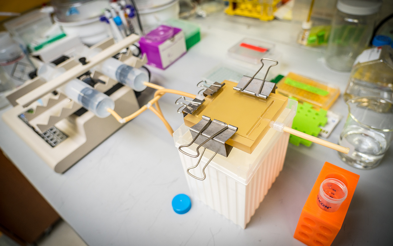
Microfluidic device sorts and separates less “sticky” cancer cells from their more sticky counterparts in the same tumor. Photo by David Baillot/UC San Diego Jacobs School of Engineering
Their work addresses a longstanding problem in the field of cancer research: it has been difficult to find biological markers to universally identify and select the most aggressive cells in tumors. This study may provide a much-needed physical marker that identifies highly metastatic cells within a heterogeneous tumor cell population.
“This new device could be the first step to better assess how likely tumor recurrence is,” said Adam Engler, bioengineering professor at the UC San Diego Jacobs School of Engineering and senior author of the study. “Patients with few of these aggressive cells lying dormant in their surrounding tissue may be less likely to see a tumor reoccur 5, 10, or 20 years later.” Engler noted that by knowing a patient’s risk, follow-up treatments could be better tailored to the individual.
The device that Engler’s team built consists of a microfluidic chamber coated with an adhesive protein. Cancer cells are placed in the chamber and after they adhere, a fluid is pushed through to detach cells. The faster the fluid moves, the higher the shear stress that the cells experience. The team can isolate cells that detach at specific shear stresses and analyze them. Cells collected at lower shear stress are weakly adherent, while those collected at higher shear stresses are strongly adherent.
Their analysis led the team to another critical finding: weakly adherent cells have a unique genetic signature that identifies them and enables them to migrate and invade faster. Comparing this signature to thousands of patients in the Cancer Genome Atlas (TCGA) database, researchers found that patients with tumors high in this “weakly adherent signature” experienced tumor recurrence occurred earlier and more frequently.
Building on these findings, Engler and his team hope to “prime” tumors with weakly adherent cells to see if they indeed metastasize faster and more frequently.
“If our mouse model shows that these cells indeed reduce cancer-free survival times, it will pave the way for substantial prognostic studies in humans with these types of solid tumors,” said first author Pranjali Beri, a bioengineering Ph.D. student in Engler’s lab. Beri also noted that nearly any solid tumor should exhibit this physical marker, and the team has so far tested cells from breast, prostate, and lung tumors.
In the future, the team hopes that clinicians will use this microfluidic device to examine tumor biopsies to estimate the likelihood of metastasis and adjust treatment at earlier disease stages.
Engler’s clinical collaborator, Dr. Anne Wallace, director of the Comprehensive Breast Health Center at UC San Diego Health who will provide patient samples for follow-up studies, concurred and confirmed this approach. “Many cancers that we see in the clinic, such as ductal carcinoma in situ or DCIS, remain dormant for years. It is nearly impossible for us to predict which fraction of that population will reoccur,” she noted. The team’s device could be the first to address these hard to predict recurrences.
Source: UC San Diego