
Approach seeks to expand understanding of biochemical activities common to all animals, plants and fungi.
Scientist are now combining recent advances in evolutionary analysis and deep learning to build three-dimensional models of how most proteins in eukaryotes interact. (Eukaryotes are organisms whose cells have a membrane-bound nucleus to hold genetic materials.)
The research effort has implications for understanding the biochemical processes that are common to all animals, plants, and fungi. The open-access work appears in Science.
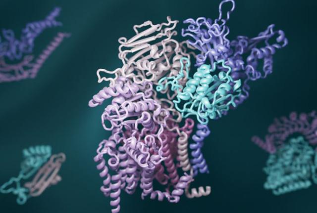
3D computed structural modeling of protein interactions is made possible by deeo learning and evolutionary analysis. Image credit: Ian Haydon, UW Medicine Institute for Protein Design
As part of a multi-institutional collaboration, the lab of David Baker at the UW Medicine Institute for Protein Design helped guide this new development.
“To really understand the cellular conditions that give rise to health and disease, it’s essential to know how different proteins in a cell work together,” Baker said. “In this paper, we provide detailed information on protein interactions for nearly every core process in eukaryotic cells. This includes over a hundred interactions that have never been seen before.”
Proteins, the workhorses of all cells, rarely act alone. Different proteins often must fit together to form precise complexes that carry out specific tasks. These can include reading genes, digesting nutrients, and responding to signals from neighboring cells and the outside world. When protein complexes malfunction, disease can result.
“This work shows that deep learning can now generate real insights into decades-old questions in biology — not just what a particular protein looks like, but also which proteins come together to interact,” said senior author Qian Cong, assistant professor of biophysics at the University of Texas Southwestern Medical Center.
To exhaustively map the interactions that give rise to protein complexes, a team of structural biologists from UW Medicine, University of Texas Southwestern Medical Center, Harvard University, and several other institutions examined all known gene sequences in yeast. Using advanced statistical analyses, they identified pairs of genes that naturally acquire mutations in a linked fashion. They reasoned that such shared mutations are a sign that the proteins the genes encode must physically interact.
The researchers also used new deep-learning software to model the three-dimensional shapes of these interacting proteins. RoseTTAFold, invented at UW Medicine, and AlphaFold, invented by the Alphabet subsidiary DeepMind, were both tapped to generate hundreds of detailed pictures of protein complexes.
“As computer methods become more powerful, it is easier than ever to generate large amounts of scientific data, but to make sense of it still requires scientific experts,” said Baker, who is a University of Washington School of Medicine professor of biochemistry. “So we recruited a village of expert biologists to interpret our 3D protein models. This is community science at its best.”
The hundreds of newly identified protein complexes provide rich insights into how cells function. For example, one complex contains the protein RAD51, which is known to play a key role in DNA repair and cancer progression in humans. Another includes the poorly understood enzyme glycosylphosphatidylinositol transamidase, which has been implicated in neurodevelopmental disorders and cancer in humans. Understanding how these and other proteins interact may open the door to the development of new medications for a wide range of health disorders.
The protein structures generated in this work are available to download from the ModelArchive. The researchers thank and remember the late John Westbrook at the Protein Data Bank for his support in establishing formats and software code to allow efficient deposition of the models into the archive. The Science paper reporting the results was in preparation at the time of Westbrook’s death.
Source: University of Washington