
Filling bone defects after removal оsteomyelitis sequesters remains an important issue. Various compression devices used for this purpose have shown satisfactory results in some cases. However, such treatment requires much time and should be carried out in a hospital environment.
A cell-based treatment modality for osteomyelitis has been developed based on the administration of cells placed in plasma clot. Cell-laden plasma clot is implanted in the sterile bone defect to promote bone regeneration. Such treatment has been found to be safe and well tolerated. A total of 18 patients with osteomyelitis aged from 18 to 61 years were treated with cell transplants:
- 9 patients with osteomyelitis of femur;
- 7 patients with osteomyelitis of the shin bone (tibia);
- 1 patient with osteomyelitis of the sacroiliac articulation (pyogenic sacroiliitis);
- 1 patient with osteomyelitis of sternum.
X-ray examination showed that osteomyelitic cavities were reduced (signs of bone healing), and no signs of disease relapse were observed in 15 patients, suggesting that cell transplantation technology could be very effective for treating patients with osteomyelitis.
Examples.
A 40 year-old female patient underwent metal osteosynthesis due to fracture of the left femur, with subsequent development of posttraumatic osteomyelitis. Metal plate was removed and a framed structure was applied, however a necrotizing 15 sm focus (sequestration) developed in the shaft of femur (Figure 1, left image). This focus was resected (Figure 1, right image) and bone defect was closed with cell implant. Subsequent repeated cell transplantation procedures resulted in the secondary clavus formation (Figure 2), which enabled the patient to walk with orthosis.

Figure 1.
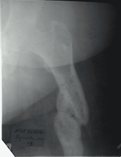
Figure 2.
A 18 year-old male patient P was subjected to sequestrectomy to resolve hematogenous osteomyelitis of the right femur, which led to the formation of a cavity with fistula within the bone defect (Figure 3). After several cell implantations, the bone defect closed (Figure 4), the patient could walk without aid and had no complains.
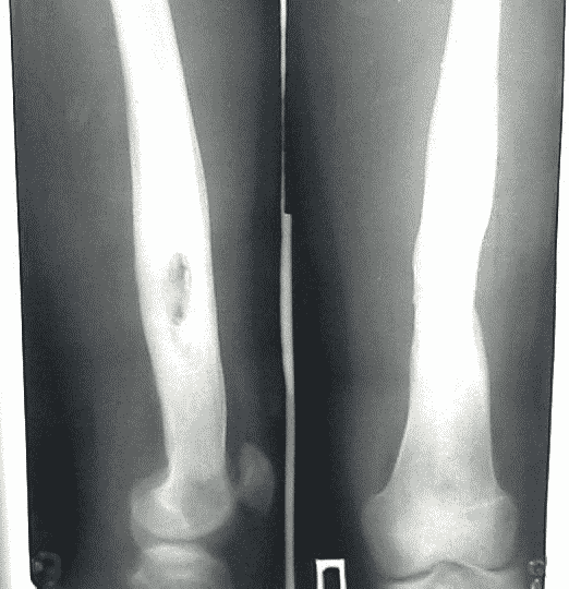
Figure 3.
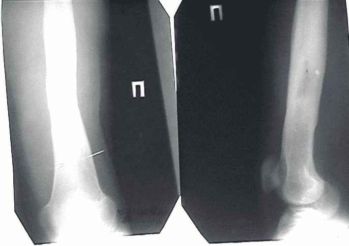
Figure 4.