
Mammals have bipartite blood circulation, i.e. pulmonary and systemic circulation. The heart is also divided: each circulatory system has an atrium and a ventricle, with the left and right halves of the heart differing in form and function. Scientists from the Max Planck Institute for Heart and Lung Research in Bad Nauheim have now found that the heart of terrestrial vertebrates evolved from the heart of primaeval fish. Ironically, an animal that does not have this divided heart, the zebrafish, helped the researchers to arrive at this conclusion. The findings may be relevant to the therapy of congenital heart defects.

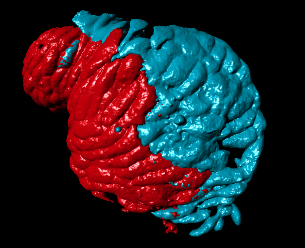
3D reconstruction of microscopic images of the fish heart during embryonic development. The image shows the atrium with asymmetric gene activity. Compartments with meis2b activity are coloured blue. Credit: MPI for heart and lung research
One of the most important preconditions for the development of terrestrial vertebrates in the course of evolution was the formation of the two halves of the circulatory system. Aquatic ancestors invariably had a single blood circulation system. In the mammal organism, however, there are two completely separate systems, the systemic and the pulmonary system. Systemic circulation supplies the body with blood, while pulmonary circulation pumps spent blood into the lungs for oxygenation. This is why the heart has a pair of atriums and a pair of ventricles. However, the left half of the heart differs considerably from the right half.
It is still largely unclear how the two circulatory systems develop. Through their studies of zebrafish, scientists at the Max Planck Institute for Heart and Lung Research have found that the mammalian heart is an evolutionary development of the fish heart. “Although the latter consists of only one ventricle and one atrium, the activity of different genes indicate a division into two parts, especially in the atrium”, says Almary Guerra, lead author of the study.
Different gene activity
The researchers concentrated particularly on two genes, meis2b and pitx2c. “In the zebrafish, both genes are highly active in the atrium. What was especially exciting was that the genes were only particularly active in a group of progenitor cells, from which the left part of the atrium later develops”, says Guerra. Based on this, the Max Planck researchers concluded that there are already genetic differences within the fish atrium, which are also found in the mammal heart. “A series of genes that are crucial to the properties of the left atrium of the human heart are also active only on the left side of the atrium in zebrafish”, says Sven Reischauer, Group Leader at the Max Planck Institute. For example, defects in some of these genes can lead to what are known as septal defects, in which the left and right halves of the heart are not completely separated.
To examine whether a genetic defect also leads to development problems in the heart of the zebrafish, the researchers turned off the meis2b gene by genetic manipulation. The fact is, development of the zebrafish heart is flawed without meis2b. “The shape and size of the atrium and the transmission of stimuli is impaired. A similar symptom is also recognized in people with a defect in the genes corresponding to meis2b and pitx2c”, says Reischauer.
In their study, the Max Planck researchers were able, for the first time, to detect the formation of compartments in the atrium of the fish heart, which is comparable with the formation of the two atria in the terrestrial vertebrates. “Next, we will explore further details leading to the asymmetry of the atrium in fish”, says Reischauer. The aim of the scientists in Bad Nauheim is to also facilitate better understanding of congenital heart disease and ultimately to create the basis for their therapy.
Source: MPG