
For patients diagnosed with a certain type of brain cancer called glioblastoma, long-term survival is rare. Surgery can remove tumors visible to the surgeon’s eye. It cannot, however, take out the diseased tendrils of tumor cells that extend beyond what the doctor sees. While current immunotherapy drugs are effective for several types of cancers, they do not work on glioblastomas because the disease blocks white blood cells from entering the tumor.
Researchers at the University of California San Diego, however, figured out a way to combine FDA-approved ultrasound with engineered glass particles to boost the effectiveness of immunotherapy in glioblastomas. Their findings were published in Advanced Therapeutics.

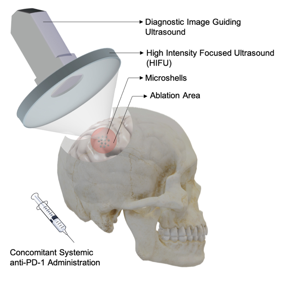
Figure 1. Schematic of microshells enhancing HIFU in a potential glioblastoma clinical setting. Glioblastoma tumors initially suppress the immune system. Microshells enhanced image-guided HIFU to blow up the tumors and release tumor antigens. Simultaneous systemic blocking of PD-1, the break on T cells re-awakens the immune system to establish tumor immunity. Figure by James Wang
Professor of Chemistry and Biochemistry Andrew Kummel, his former doctoral candidate and lead author of the paper James Wang and their collaborators, including Clark Chen at the University of Minnesota Medical School, developed this highly effective approach for taking on glioblastoma. They created hollowed particles, called microshells, made of silica (glass) and filled them with near body-temperature fluorocarbon liquid—a type of highly volatile and inert substance that is FDA-approved as an ultrasound contrast agent. The scientists injected the microshells into glioblastomas in mice and used high intensity focused ultrasound (HIFU) to blow up the shells inside the tumor. The shear force generated by the explosions ruptured the cancer cells to release tumor proteins.
“The silica shells are like micro-sized Christmas tree ornaments,” explained Kummel. “The HIFU ultrasound is like a cat paw that breaks them open. The liquid inside turns to gas when the nanoshells break, thereby rupturing the cancer cells.”
Kummel added that the blast releases tumor proteins which attract white blood cells to the tumor. “When combined with the immunotherapy that is currently approved for patient use, these released proteins train the immune system to combat the tumor,” he said.
Chen, co-author on the paper, agreed. “Without white blood cells inside the tumor, immunotherapy does not work. The trick is to find ways to help the white blood cells get into the tumor.”
That trick, according to Chen, involves rupturing the cancer cells at body temperature—something they could do with HIFU—because higher temperatures compromise the effectiveness of white blood cells to fight off the tumor.
Kummel commented that the study may pave the way for rapid use in human clinical trials because of HIFU—even without microshells. While the microshells would be more directly effective, getting them approved would take years. But HIFU combined with a currently FDA-approved drug could help glioblastoma patients now.
Originally, the microshells were developed by UC San Diego’s Professor of Chemistry and Biochemistry William Trogler and Surgical Oncologist Sarah Blair of UC San Diego Health, Moores Cancer Center, for marking breast tumors for surgical removal. Kummel and his team, however, are the first to apply the microshells in a way to bolster the effectiveness of immunotherapy against glioblastoma.
Source: UC San Diego